

Information, opinions and surveys from Moscow
Conclusion
(You can enlarge by clicking on it)
Translate:
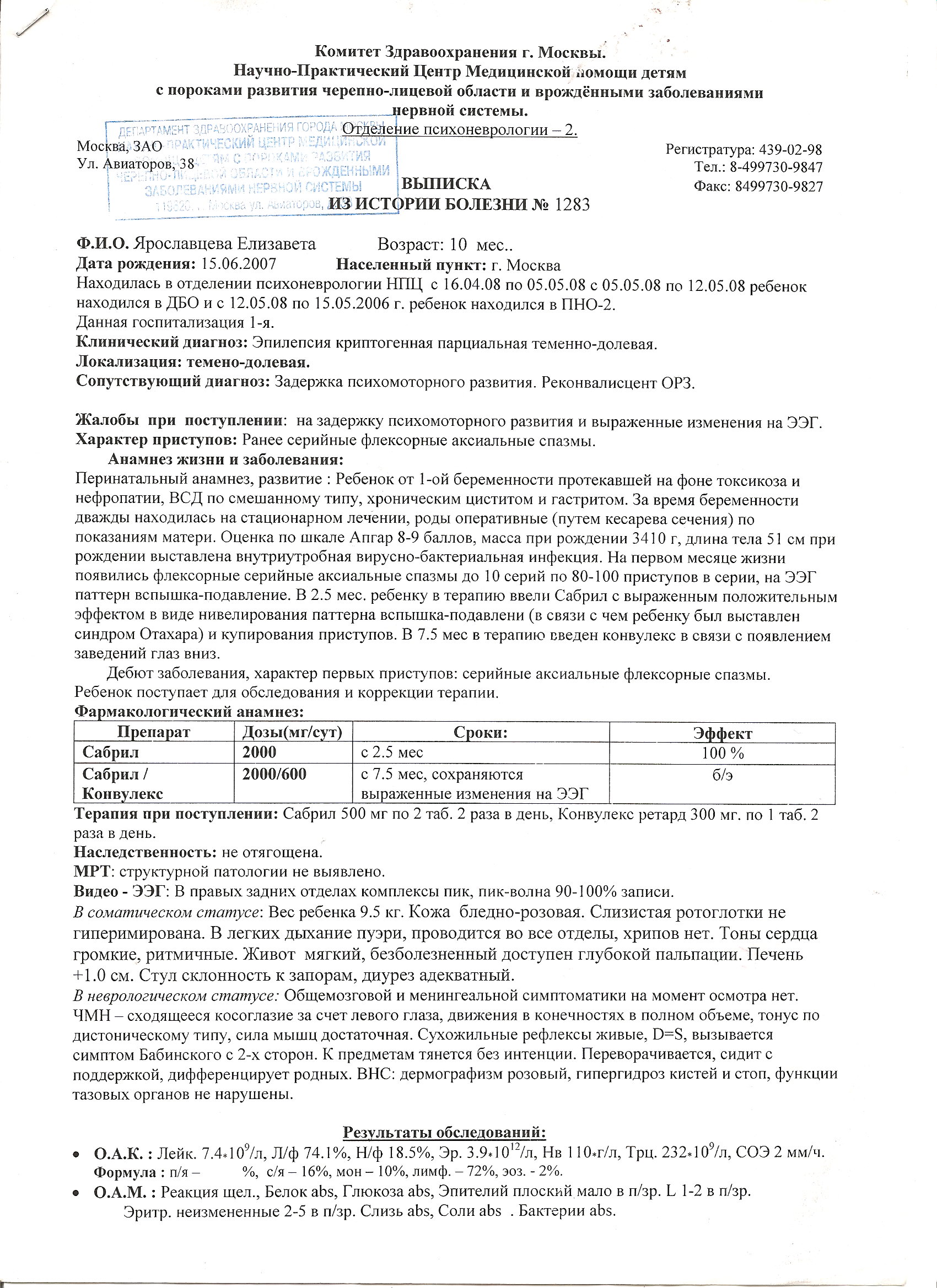
Federal State Establishment “Moscow SRI of Pediatrics and Children’s Surgery” RusMedTechnology
Department of Neuropsychiatry and Epileptology
Children’s scientific-practical anti-seizure center
125412 Moscow, Taldomskaya 2
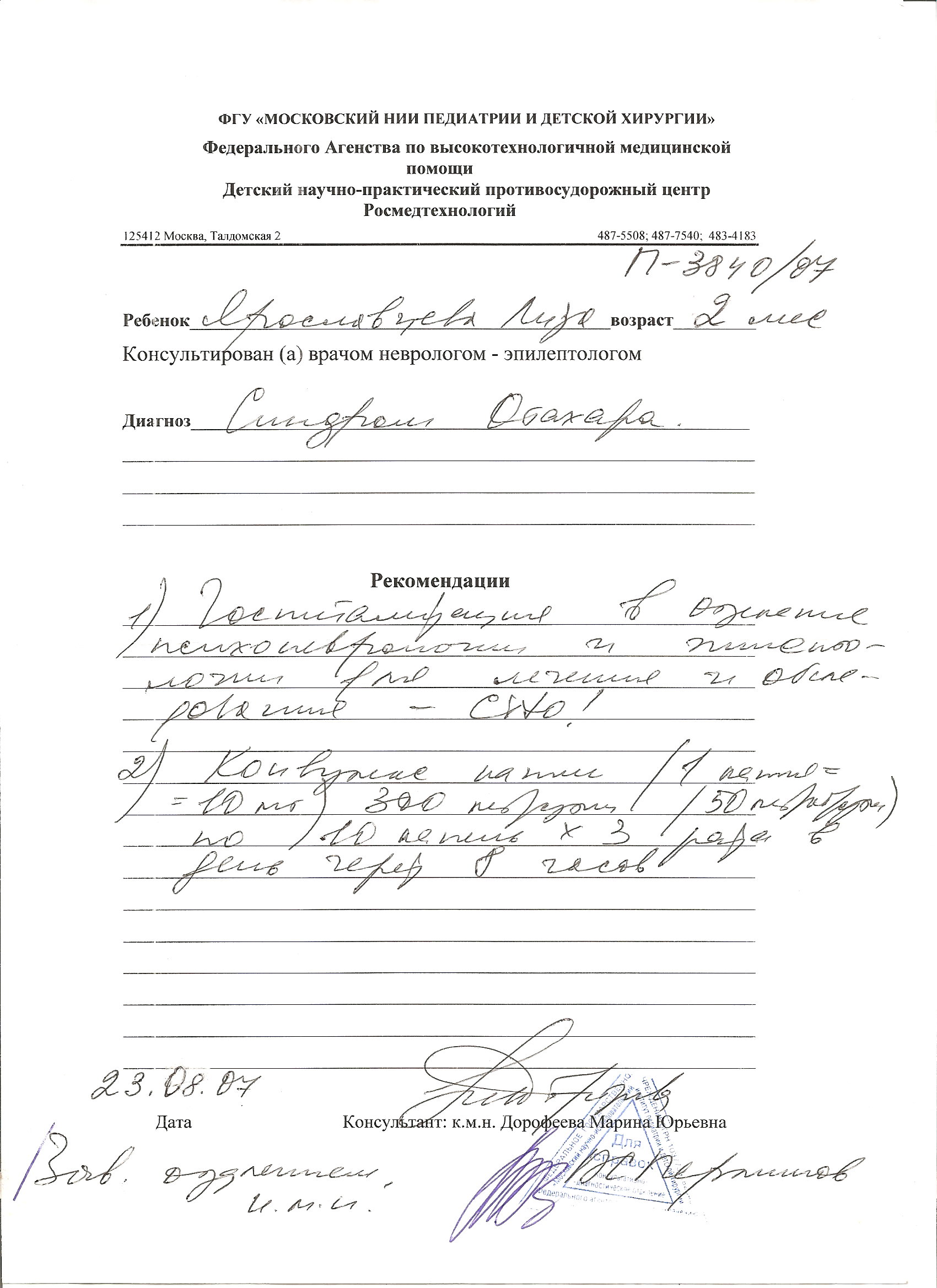
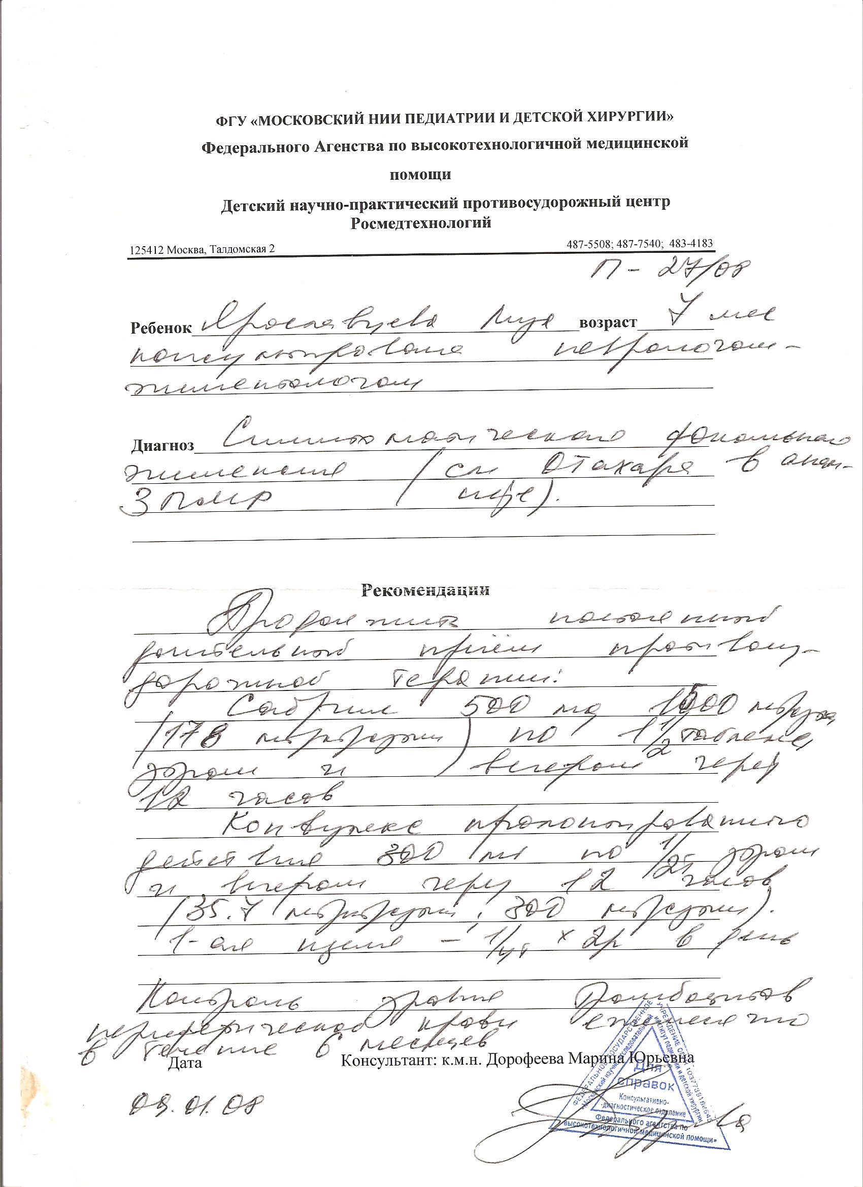
Child Yaroslavtseva Elizaveta, 1 year and 5 months old (AK# 821/08) has
been observed in the Children’s scientific-practical anti-seizure
center of Moscow SRI of Pediatrics and Children’s Surgery
RusMedTechnology since the age of 2 months.Department of Neuropsychiatry and Epileptology
Children’s scientific-practical anti-seizure center
125412 Moscow, Taldomskaya 2
Diagnosis: Symptomatic Fractional Epilepsy, resistant course. Ohtahara Syndrome in anamnesis. Psychomotor development delay.
Ohtahara Syndrome was diagnosed at the age of 2 months, which was preceded by tonic eye abduction to left and up. Seizures disappeared at the age of 3 months and 2 weeks as the result of treatment with Sabril (Vigabatrin) 2000 mg/day. At the age of 7 months the series of eye abduction repeated 10-30 times per series up to 20 series a day. Convulex Retard was added to the therapy without positive result. During EEG video monitoring prolonged epileptic activity was registered in occipital region with accent to the right. The seizures continued during hospitalization. Frizium (klobazam) 15 mg/day was added to the therapy with clear positive effect, dose of Sabril was lowered to 1500 mg/day. The seizures repeated at the age of 11 months (05/05/2008) as tonic myotonia with eye abduction up to 5 seizures, 40 seconds in length up to 2 times a day.
Currently the child is being given anti-seizure prescriptions: Convulex “300 mg” 1 pill twice a day with 12 hours interval (600 mg/day, 50 mg/kg/day), Sabril “500 mg” 1½ pill twice a day with 12 hours interval (1500 mg/day, 125 mg/kg/day) and Frizium “10 mg” 1 pill twice a day (20 mg/day; 1,6 mg/kg/day).
Magnetic-resonance brain tomography August 22, 2007: Focal Cortical Dysplasia in the right occipital region.
The examination to determine the possibility of surgical treatment of epilepsy in Epileptology Center of Bielefeld (Germany) is recommended.
Date: November 14 2008
Doctor: M. Y. Dorofeeva
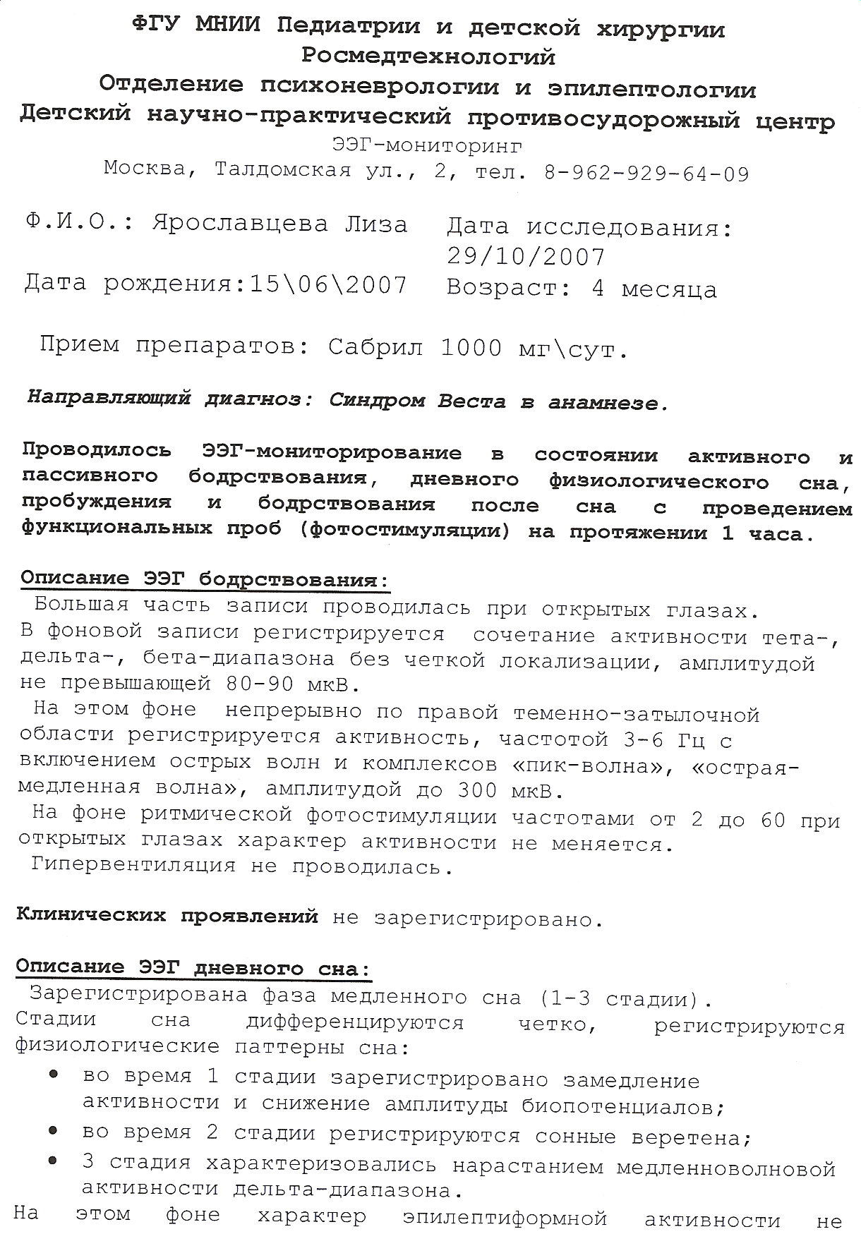
Survey EEG
(You can enlarge by clicking on it)
Translate:
Federal State Establishment “Moscow SRI of Pediatrics and Children’s Surgery” RusMedTechnology
Department of Neuropsychiatry and Epileptology
Children’s scientific-practical anti-seizure center
125412 Moscow, Taldomskaya 2. Tel. 8-962-929-64-09
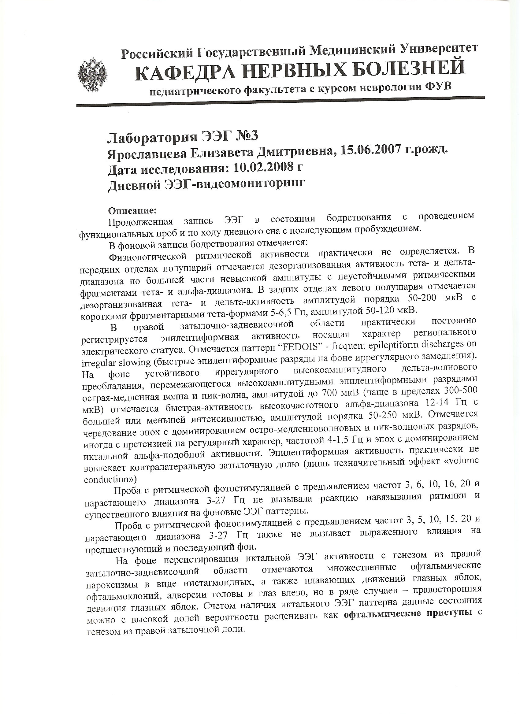
Name: Yaroslavtseva Liza
Date of Birth: June 15, 2007 Age 4 months
Date of research October 29, 2007
Prescriptions: Sabril 1000 mg/day
Diagnosis West Syndrome in anamnesis
EEG monitoring was held during active and passive wakefulness, during day physiological sleep, awakening and activity after sleep with functional test (photo stimulation) during 1 hour.
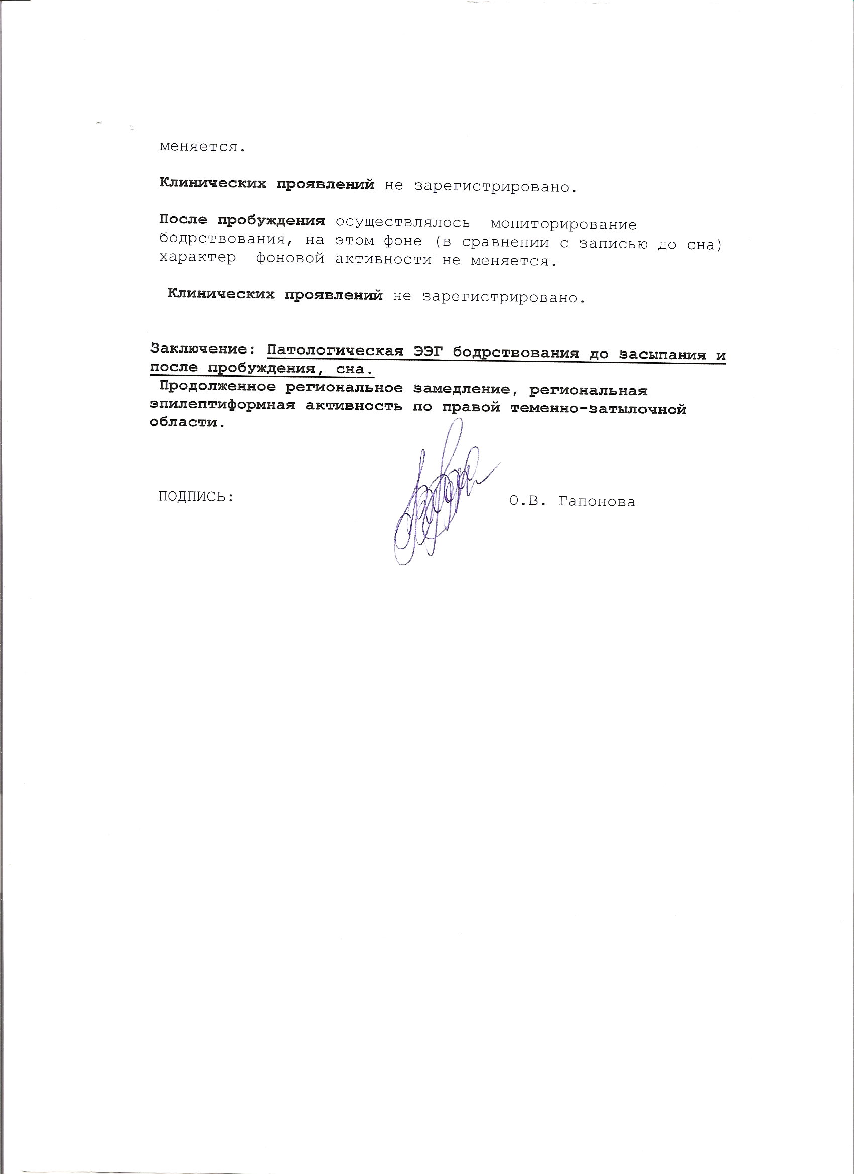
Description of EEG wakefulness:
The majority of recording took place with open eyes.
Baseline recording registered combination of activity theta-, delta-, beta-range without distinct localization with amplitude not exceeding 80-90 mkB.
In this baseline continuous activity in right parietooccipital area has been registered with frequency of 3-6 Hz with sharp waves and “top-wave” complex inclusions, “sharp-slow wave”, with amplitude up to 300 mkB.
In baseline of rhythmical photo stimulation with frequency from 2 to 60 with open eyes the activity nature did not change.
Clinical presentation did not register.
Description of EEG of day sleep:
The phase of slow wave sleep has been registered (1-3 stages).
The stages of sleep differentiate distinctively; physiological patterns of sleep have been registered:
-during stage 1 slowing-down has been registered;
-during stage 2 sleep spindles has been registered;
-stage 3 has been characterized with augmentation of slow wave activity of delta range.
In this baseline the nature of epileptoid activity did not change.
Clinical presentation did not register.
After awakening the monitoring of wakefulness was held, in this baseline the nature of activity did not change in comparison with monitoring before sleep.
Clinical presentation did not register.
Conclusion: Pathological EEG of wakefulness before sleep and after awakening, and during sleep.
Continuous regional slowing down, regional epileptical activity in right parietooccipital area.
Signature: O.V. Gaponova
______________________________Date of Birth: June 15, 2007 Age 4 months
Date of research October 29, 2007
Prescriptions: Sabril 1000 mg/day
Diagnosis West Syndrome in anamnesis
EEG monitoring was held during active and passive wakefulness, during day physiological sleep, awakening and activity after sleep with functional test (photo stimulation) during 1 hour.
Description of EEG wakefulness:
The majority of recording took place with open eyes.
Baseline recording registered combination of activity theta-, delta-, beta-range without distinct localization with amplitude not exceeding 80-90 mkB.
In this baseline continuous activity in right parietooccipital area has been registered with frequency of 3-6 Hz with sharp waves and “top-wave” complex inclusions, “sharp-slow wave”, with amplitude up to 300 mkB.
In baseline of rhythmical photo stimulation with frequency from 2 to 60 with open eyes the activity nature did not change.
Clinical presentation did not register.
Description of EEG of day sleep:
The phase of slow wave sleep has been registered (1-3 stages).
The stages of sleep differentiate distinctively; physiological patterns of sleep have been registered:
-during stage 1 slowing-down has been registered;
-during stage 2 sleep spindles has been registered;
-stage 3 has been characterized with augmentation of slow wave activity of delta range.
In this baseline the nature of epileptoid activity did not change.
Clinical presentation did not register.
After awakening the monitoring of wakefulness was held, in this baseline the nature of activity did not change in comparison with monitoring before sleep.
Clinical presentation did not register.
Conclusion: Pathological EEG of wakefulness before sleep and after awakening, and during sleep.
Continuous regional slowing down, regional epileptical activity in right parietooccipital area.
Signature: O.V. Gaponova
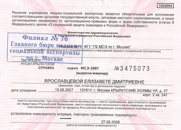
Information on disability
(You can enlarge by clicking on it)
______________________________
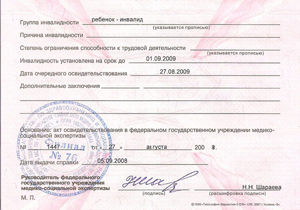
Information on disability reverse side
(You can enlarge by clicking on it)
______________________________
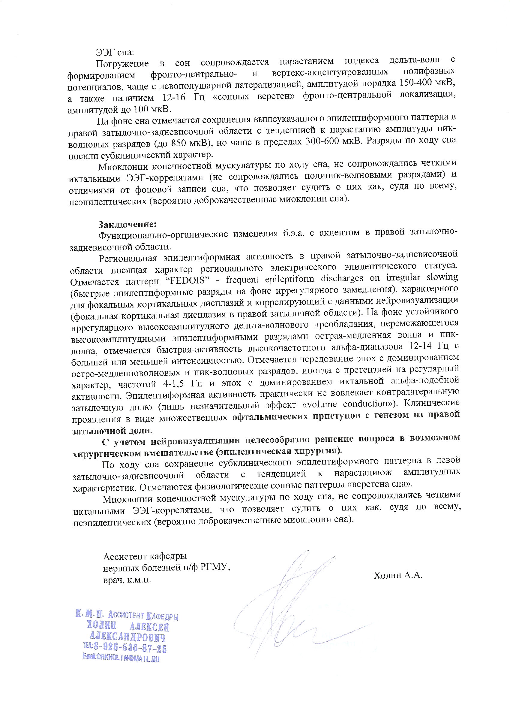
Videomonitoring EEG
(You can enlarge by clicking on it)
______________________________
Consultation and diagnosis
(You can enlarge by clicking on it)
______________________________
The diagnosis and recommendations
(You can enlarge by clicking on it)
______________________________
An extract from the history of the disease
(You can enlarge by clicking on it)